Canker Sore vs Oral Cancer: Key Differences and Warning Signs
A painful spot inside your mouth appears, and a wave of anxiety follows. Is it just a common canker sore, or could it be something more serious, like oral cancer? This question is a source of significant worry for many people. While the vast majority of mouth sores are benign and self-limiting, understanding the distinct characteristics that separate a harmless canker sore from a potential oral cancer lesion is crucial for your health and peace of mind. This guide provides a detailed, side by side comparison to help you recognize the typical features of each condition, know when to seek professional medical evaluation, and understand the importance of early detection.
Understanding Canker Sores: The Common Mouth Ulcer
Canker sores, medically known as aphthous ulcers, are small, shallow, non contagious lesions that develop on the soft tissues inside your mouth. They are incredibly common, affecting a large portion of the population at some point in their lives. These sores are not caused by a virus (unlike cold sores) and cannot be spread through kissing or sharing utensils. The exact cause of canker sores is not fully understood, but they are often linked to a combination of factors that trigger an immune system response in the delicate oral mucosa.
Common triggers include minor mouth injuries from dental work, aggressive brushing, or accidental bites. Food sensitivities, particularly to acidic foods like citrus fruits, tomatoes, or chocolate, can also precipitate an outbreak. Nutritional deficiencies, especially in vitamin B12, zinc, folate, or iron, are another contributing factor. Furthermore, emotional stress, hormonal shifts, and certain underlying health conditions, such as Crohn’s disease or celiac disease, can make individuals more susceptible. It is important to note that canker sores are not precancerous and do not increase your risk of developing oral cancer.
Recognizing the Signs of Oral Cancer
Oral cancer, which includes cancers of the lips, tongue, cheeks, floor of the mouth, hard and soft palate, sinuses, and pharynx, is a serious medical condition. It occurs when cells in the oral cavity develop mutations in their DNA, leading to uncontrolled growth and the formation of a malignant tumor. The primary risk factor for oral cancer is tobacco use in any form, including cigarettes, cigars, pipes, and chewing tobacco. Heavy alcohol consumption significantly increases risk, and the combination of tobacco and alcohol is particularly dangerous. Other risk factors include infection with the human papillomavirus (HPV), specifically HPV16, prolonged sun exposure to the lips, a diet low in fruits and vegetables, and a weakened immune system.
Oral cancer lesions can manifest in various ways, and they often do not cause pain in their earliest, most treatable stages. This silent progression is why regular self examinations and dental checkups are vital. Unlike canker sores, which follow a predictable cycle of appearing, peaking in pain, and healing, cancerous growths tend to persist and evolve. They may start as a subtle change in tissue color or texture that gradually becomes more pronounced. Because early detection dramatically improves treatment outcomes and survival rates, knowing the warning signs is a critical component of oral health awareness.
Canker Sore vs Cancer: A Direct Symptom Comparison
Distinguishing between a canker sore and a potentially cancerous lesion hinges on observing several key characteristics: appearance, sensation, duration, and location. The following comparison outlines the typical features of each. Use this as a guide, not a definitive diagnosis. Any sore or abnormality that causes concern warrants a professional evaluation by a dentist or doctor.
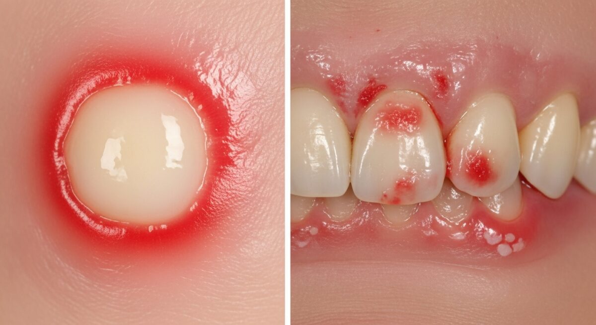
Appearance and Texture: A typical canker sore is round or oval with a well defined border. It has a white or yellowish center (a pseudomembrane) surrounded by a bright red halo of inflammation. The sore is shallow and sits on the surface of the mucosa. In contrast, an oral cancer lesion may appear as a red patch (erythroplakia), a white patch (leukoplakia) that cannot be scraped off, or a mixed red and white patch. It may also present as a lump, thickening, or rough spot. The borders are often irregular, ragged, or poorly defined. The lesion may be raised, crusted, or have an ulcerated center that feels hard to the touch.
Sensation and Pain: Canker sores are famously painful. The pain is often disproportionate to their size, especially when eating, drinking, or talking. The discomfort usually begins a day or two before the ulcer becomes fully visible and peaks in the first few days. Oral cancer lesions, particularly in early stages, are frequently painless. As the cancer advances, it may cause a persistent sore throat, a feeling that something is caught in the throat, numbness in the mouth or lips, pain in the ear, or pain when swallowing. Pain, if it occurs, is typically a later symptom.
Duration and Healing: This is one of the most critical differentiators. A canker sore follows a self limiting course. It will usually heal completely within 10 to 14 days without any medical intervention. Oral cancer sores do not heal. They persist for weeks, months, or continue to grow larger. A lesion that remains unchanged or worsens beyond a two week period must be examined by a healthcare professional.
Common Locations: Canker sores appear exclusively on the movable, non keratinized tissues inside the mouth. This includes the inside of the lips and cheeks, the floor of the mouth, under the tongue, and the soft palate. They do not occur on the keratinized surfaces like the gums, the hard palate, or the top surface of the tongue. Oral cancer can develop anywhere in the oral cavity, but common sites include the sides and underside of the tongue, the floor of the mouth, the lips, and the area at the back of the mouth/top of the throat (oropharynx). Sores on the gums, hard palate, or top of the tongue are less likely to be canker sores and should be assessed.
When to See a Doctor or Dentist: Red Flags
You should schedule an appointment with your dentist or primary care physician if you notice any of the following warning signs. Do not wait for the symptom to become painful. Early evaluation is key.
- A sore, lump, or patch inside your mouth that does not heal within two weeks.
- A persistent white, red, or speckled patch that does not rub off.
- A growth, lump, or thickening of the skin or lining inside your mouth.
- Unexplained bleeding in the mouth.
- Persistent numbness, loss of feeling, or pain/tenderness in any area of the face, mouth, or neck.
- Difficulty chewing, swallowing, speaking, or moving your jaw or tongue.
- A change in your voice or a persistent feeling that something is caught in your throat.
- Ear pain without hearing loss.
- A dramatic change in the way your teeth or dentures fit together.
- Unexplained weight loss.
During your visit, the professional will conduct a visual and tactile examination of your mouth, head, and neck. If a suspicious area is found, the next step is typically a biopsy. This is a minor procedure where a small sample of tissue is removed and sent to a pathology lab for analysis. A biopsy is the only definitive way to diagnose or rule out oral cancer. It is a straightforward and crucial step that provides a clear answer and guides any necessary treatment planning.
Diagnosis, Treatment, and the Role of Regular Screenings
The diagnostic pathways for canker sores and oral cancer are fundamentally different. For recurrent or severe canker sores, a doctor may review your medical history, diet, and lifestyle to identify triggers. Blood tests might be ordered to check for nutritional deficiencies or underlying systemic conditions. Treatment focuses on symptom management and may include topical anesthetics, antimicrobial mouth rinses, or corticosteroid pastes to reduce inflammation and pain.
For oral cancer, diagnosis begins with the biopsy. If cancer is confirmed, further imaging tests, such as CT scans, MRIs, or PET scans, are used to determine the stage of the cancer, which indicates its size and whether it has spread. Treatment is multidisciplinary and may involve surgery, radiation therapy, chemotherapy, targeted drug therapy, or immunotherapy, often in combination. The treatment plan is highly individualized based on the cancer’s location, stage, and the patient’s overall health.
This underscores the irreplaceable value of regular oral cancer screenings. During a routine dental checkup, your dentist or dental hygienist performs a visual and physical examination of your entire oral cavity, looking for any abnormalities. These screenings are quick, painless, and can detect early signs of cancer before you even notice symptoms. Adults should have an oral cancer screening at least once a year, and more frequently if they have risk factors.
Frequently Asked Questions
Can a canker sore turn into cancer?
No. Canker sores are benign lesions with no known potential to become cancerous. They are a separate condition with a different etiology from oral cancer.
How can I tell if a lump in my mouth is serious?
Any new lump that persists for more than two weeks, is hard or fixed in place, grows in size, or is accompanied by other symptoms like numbness or bleeding should be evaluated by a professional immediately. A soft, movable lump that comes and goes quickly is less concerning.
Are white patches always cancer?
No. Many white patches, such as those from cheek biting, friction, or thrush (a fungal infection), are benign. However, a white patch that is persistent, cannot be wiped away, and has a rough or hardened texture (leukoplakia) is considered a precancerous condition and requires biopsy.
What is the survival rate for oral cancer?
Survival rates are highly dependent on the stage at diagnosis. The five year survival rate for localized oral cancer (cancer that has not spread) is around 85%. This drops significantly if the cancer has spread to distant parts of the body. This statistic powerfully highlights why early detection through self exams and professional screenings is so critical.
How often should I check my own mouth for signs of cancer?
Perform a self examination once a month. In good light, look and feel inside your lips, cheeks, gums, the roof and floor of your mouth, and all surfaces of your tongue. Check your neck and jaw for lumps or swellings. Familiarity with your normal anatomy makes it easier to spot changes.
Empowering yourself with knowledge about the differences between common canker sores and oral cancer is a vital step in taking charge of your oral health. While most mouth sores are harmless, adopting a proactive approach that includes regular self checks, routine dental visits for professional screenings, and prompt medical consultation for any persistent abnormality is the best strategy for early detection and successful outcomes. Your vigilance, combined with professional care, forms the strongest defense against oral health concerns.