Skin Cancer Pictures: A Visual Guide to Identifying Suspicious Spots
Noticing a new or changing spot on your skin can be worrying. While online skin cancer pictures can be a helpful starting point for visual learning, they are not a substitute for a professional medical diagnosis. This guide aims to bridge the gap between seeing an image and understanding what to do next. We will walk you through the visual hallmarks of major skin cancer types, explain the critical limits of self diagnosis, and outline the clear steps to take if you see something concerning.
Understanding the Limits of Visual Comparisons
Before diving into descriptions and comparisons, it is crucial to establish a fundamental rule. Looking at skin cancer pictures online is an educational tool, not a diagnostic one. Many benign skin conditions, such as seborrheic keratoses, harmless moles, or skin tags, can resemble cancerous growths to the untrained eye. Conversely, some dangerous melanomas can look deceptively innocent. Relying solely on image matching can lead to unnecessary anxiety or, more dangerously, a false sense of security. The true purpose of reviewing these pictures is to build awareness of the ABCDEs and other warning signs, so you know when it is time to seek expert evaluation. Your most powerful tool is a combination of this knowledge and a commitment to regular self exams and professional skin checks.
The ABCDE Framework: Your First Filter
Dermatologists use the ABCDE rule as a simple, memorable guide for assessing moles and spots. This framework applies primarily to melanoma, the most dangerous form of skin cancer, but its principles are useful for general vigilance. When you examine your skin or look at reference pictures, run through this checklist. Asymmetry is the first sign. Draw an imaginary line through the middle of a spot. If the two halves do not match, it is considered asymmetrical, a warning sign. Border irregularity is next. Look for edges that are ragged, notched, scalloped, or blurred instead of smooth and even. Color variation is a major red flag. Multiple shades of brown, tan, black, and sometimes white, red, or blue within a single lesion are concerning. Diameter matters. While melanomas can be small, a spot larger than the size of a pencil eraser, about 6 millimeters across, warrants attention. Finally, evaluate evolution. Any change in size, shape, color, elevation, or any new symptom like bleeding, itching, or crusting is the most critical sign of all. A spot that is evolving demands a prompt visit to the doctor.
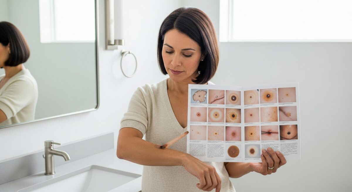
Visual Guide to Common Skin Cancer Types
Skin cancer manifests in several forms, each with distinct visual characteristics. Recognizing these differences can help you describe concerns more accurately to a healthcare provider. Below is a breakdown of the three most common types.
Basal Cell Carcinoma (BCC)
Basal cell carcinoma is the most frequently occurring skin cancer. It often appears on sun exposed areas like the face, ears, and neck. BCCs grow slowly and rarely spread, but they can cause significant local damage if left untreated. In skin cancer pictures, you might see several presentations. A nodular BCC often looks like a pearly or waxy bump, sometimes with visible blood vessels on its surface. It may resemble a translucent pimple that does not heal and might bleed or crust over. Superficial BCC can appear as a red, scaly patch, similar to eczema, but it persists and slowly expands. A morpheaform BCC is less common, presenting as a white or yellow, scar like area with poorly defined borders. These are often firmer to the touch.
Squamous Cell Carcinoma (SCC)
Squamous cell carcinoma is the second most common skin cancer. It also arises on sun exposed skin but can develop on other parts of the body, including mucous membranes. SCCs are more likely than BCCs to grow deeper and spread if neglected. Visually, SCCs often present as a firm, red nodule. They may have a rough, scaly, or crusted surface that might bleed if scratched. Sometimes they look like a persistent, thick, scaly patch that continues to grow. They can also develop from a precancerous lesion called an actinic keratosis, which appears as a dry, rough, scaly patch of skin.
Melanoma
Melanoma is less common but far more dangerous due to its potential to spread to other parts of the body. It can develop anywhere, even in areas not exposed to the sun. Melanomas often, but not always, arise from an existing mole. They are the prime example for the ABCDE rule. In addition to the ABCDEs, be aware of the “ugly duckling” sign. This refers to a mole that looks distinctly different from all the other moles on your body. It stands out in its appearance. Acral lentiginous melanoma, a type more common in people with darker skin tones, appears on palms, soles, or under nails. It may look like a dark streak under a fingernail or toenail or an irregular brown to black patch on a palm or sole.
What to Do If You Identify a Suspicious Spot
Finding a spot that matches concerning descriptions from skin cancer pictures is a signal to act, not to panic. The process is straightforward. First, do not attempt to biopsy or remove the spot yourself. This can interfere with a proper diagnosis. Schedule an appointment with a dermatologist, a doctor specializing in skin conditions. If you do not have a dermatologist, your primary care physician can perform an initial assessment and provide a referral. Before your appointment, take clear, well lit photos of the spot next to a ruler or coin for scale. This creates a visual record your doctor can use to assess change over time. During the visit, the dermatologist will perform a full body skin exam. If a lesion is suspicious, they will likely perform a biopsy. This is a simple procedure where a small sample of the tissue is removed and sent to a lab for analysis. It is the only way to definitively diagnose skin cancer.
Understanding your health insurance coverage for preventive screenings and diagnostic procedures is an important part of proactive care. For a detailed explanation of how Medicare and other plans may cover dermatology visits and skin cancer screenings, Read full article on navigating healthcare benefits for preventive services.
Prevention: Your Most Powerful Strategy
While identifying skin cancer early is crucial, preventing it in the first place is even more effective. Sun protection is non negotiable. Use a broad spectrum sunscreen with an SPF of 30 or higher every day, even on cloudy days and in winter. Apply it generously to all exposed skin and reapply every two hours, or more often if swimming or sweating. Seek shade, especially between 10 a.m. and 4 p.m. when the sun’s rays are strongest. Wear protective clothing, including a wide brimmed hat, sunglasses with UV protection, and long sleeves and pants when possible. Avoid tanning beds entirely, as they emit concentrated UV radiation that significantly increases skin cancer risk. Make monthly self exams a habit. Know your skin so you can spot changes early. Use a mirror to check hard to see areas, or ask a partner or family member for help.
Frequently Asked Questions
Can a spot be skin cancer if it does not match pictures exactly? Absolutely. Skin cancers are notoriously variable. Some may not exhibit classic ABCDE features, especially in early stages. Any new, changing, or unusual spot should be evaluated professionally.
How often should I see a dermatologist for a skin check? The recommended frequency depends on your personal risk factors. Individuals with a history of skin cancer, many atypical moles, a family history of melanoma, or a history of significant sunburn may need annual checks. Others might be advised to come every 1 3 years. Consult your doctor for personalized guidance.
Are there apps that can diagnose skin cancer from a photo? No. While some apps use algorithms to analyze images and provide risk assessments, they are not approved for diagnosis. Studies have shown they can miss melanomas. They should never replace a visit to a dermatologist.
What does skin cancer look like on darker skin tones? While less common, skin cancer can occur on all skin tones. On darker skin, it often appears in less sun exposed areas like palms, soles, under nails, or inside the mouth. Melanomas may be darker than the surrounding skin or present as dark lines under nails. SCC may appear as a hard bump or a patch that is darker than the surrounding skin. Awareness is key.
Ultimately, using skin cancer pictures as an educational reference empowers you to be an active participant in your skin health. Let them inform you, not diagnose you. Your vigilance in performing self exams, practicing consistent sun protection, and seeking professional evaluation for anything suspicious is the definitive strategy for early detection and successful treatment. When in doubt, get it checked out.