How to Spot and Treat Early Stage Skin Cancer

Finding a strange spot on your skin can send a wave of worry through anyone. That initial suspicion, however, is a powerful tool in the fight against skin cancer. When caught in its early stages, skin cancer is almost always highly treatable, often with minimally invasive procedures and excellent outcomes. Understanding what to look for and taking prompt action can make all the difference between a simple office visit and a more complex health journey. This guide empowers you with the knowledge to identify potential warning signs, understand your next steps, and navigate the path from detection to treatment with confidence.
What Is Early Stage Skin Cancer?
Early stage skin cancer refers to the initial phase of the disease when abnormal cells have formed but are confined to the top layers of the skin, typically the epidermis. At this point, the cancer has not invaded deeper tissues or spread to other parts of the body, a process known as metastasis. The term “early stage” encompasses several types of skin cancer, each with distinct characteristics, but they share the common trait of being localized and highly curable. The three most common types are basal cell carcinoma (BCC), squamous cell carcinoma (SCC), and melanoma, with melanoma being the most aggressive. Even within these types, “early stage” can have specific medical definitions. For instance, melanoma is staged from 0 to IV, with stage 0 (melanoma in situ) and stage I being considered early, where the tumor is thin and limited to the skin. Similarly, early BCC and SCC are those that are small, superficial, and show no signs of deeper invasion or spread.
The Critical Role of Early Detection
The survival statistics for skin cancer underscore the monumental importance of early detection. For melanoma, the most dangerous form, the five-year survival rate when detected at a localized, early stage is over 99%. This rate drops significantly if the cancer spreads to distant parts of the body. For non-melanoma skin cancers like BCC and SCC, early detection often means the treatment can be simple, swift, and with minimal scarring. Beyond saving lives, early detection preserves quality of life. Treatments for advanced skin cancer can be disfiguring, expensive, and involve complex surgeries, radiation, or systemic therapies. Catching it early usually allows for smaller, less invasive surgical excisions or even non-surgical options. This proactive approach transforms skin cancer from a potentially life-threatening illness into a manageable condition, emphasizing that regular self-exams and professional skin checks are not just recommendations, they are essential health practices.
Recognizing the Warning Signs: The ABCDEs and Beyond
Becoming familiar with the visual signs of early stage skin cancer is your first line of defense. The most widely taught method is the ABCDE rule for melanoma, but it also provides a good framework for being suspicious of any skin lesion.
- A is for Asymmetry: One half of the mole or spot does not match the other half.
- B is for Border: The edges are irregular, ragged, notched, or blurred.
- C is for Color: The color is not uniform. It may have differing shades of brown, black, or tan, or patches of white, red, or blue.
- D is for Diameter: While melanomas can be small, a spot larger than 6 millimeters (about the size of a pencil eraser) is a cause for concern, though smaller changing lesions should also be evaluated.
- E is for Evolving: Any change in size, shape, color, elevation, or any new symptom like bleeding, itching, or crusting.
For basal and squamous cell carcinomas, watch for different signs. These often appear on sun-exposed areas like the face, ears, neck, and hands. Basal cell carcinoma may look like a pearly or waxy bump, a flat, flesh-colored or brown scar-like lesion, or a bleeding or scabbing sore that heals and returns. Squamous cell carcinoma often presents as a firm, red nodule or a flat lesion with a scaly, crusted surface. It’s crucial to remember that skin cancer can also manifest in less common ways. A persistent, non-healing sore, a new growth, or a spot that simply looks different from all others on your body (the “ugly duckling” sign) warrants a professional opinion. Changes in existing moles are a key warning sign, and understanding broader early stage symptoms that appear on the skin can be valuable for overall health awareness.
Diagnostic Steps: From Suspicion to Confirmation
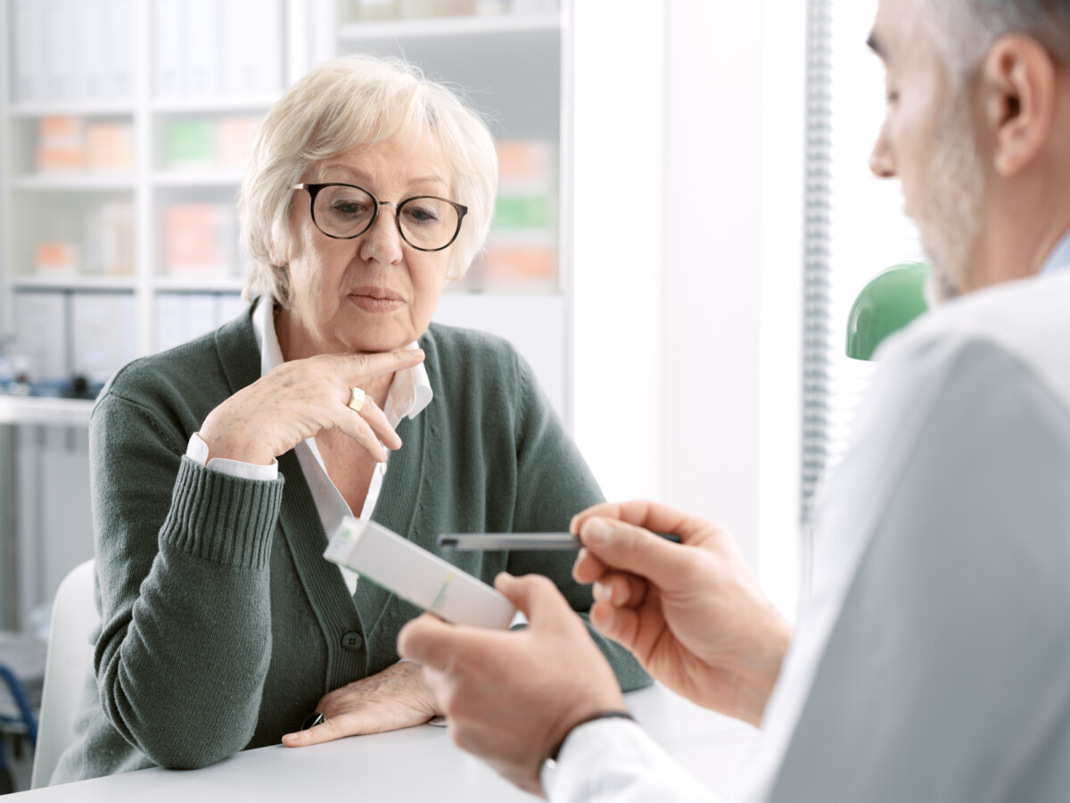
If you notice a concerning spot, the next step is to schedule an appointment with a dermatologist, a doctor who specializes in skin conditions. Do not delay. The diagnostic process typically begins with a visual examination. The dermatologist will look at the spot in question and likely perform a full-body skin check. They may use a dermatoscope, a handheld device that magnifies and lights the skin, allowing for a more detailed view of structures beneath the surface. If the lesion is suspicious, the doctor will recommend a skin biopsy to obtain a definitive diagnosis. This is a simple procedure usually done in the office under local anesthesia. The doctor removes all or part of the abnormal tissue, which is then sent to a laboratory where a pathologist examines it under a microscope. There are several types of biopsies, including shave, punch, and excisional biopsies. The choice depends on the size, location, and type of lesion suspected. This biopsy is the only way to confirm whether cells are cancerous and to identify the specific type and depth of the cancer, which guides all subsequent treatment decisions.
Treatment Options for Early Stage Lesions
The treatment plan for early stage skin cancer is highly effective and tailored to the cancer type, size, location, and the patient’s overall health. Because the cancer is confined, the primary goal is complete removal while preserving as much healthy tissue and function as possible. For many early basal and squamous cell carcinomas, the standard treatment is surgical excision. The doctor numbs the area and cuts out the tumor along with a small margin of healthy tissue to ensure all cancer cells are removed. Another common surgical technique is Mohs micrographic surgery, which is particularly valuable for cancers on the face or other cosmetically sensitive areas, or for those with ill-defined borders. In Mohs surgery, the surgeon removes thin layers of skin one at a time and examines each under a microscope during the procedure, stopping only when no cancer cells remain. This maximizes tissue preservation. For very superficial early skin cancers, non-surgical options may be suitable. These include cryotherapy (freezing with liquid nitrogen), topical medications (creams or gels applied to the skin), curettage and electrodesiccation (scraping away the cancer and sealing the wound with an electric needle), and photodynamic therapy (using a light-activated drug). For early melanoma (melanoma in situ), wide local excision, removing the tumor with a larger margin of healthy skin, is the standard treatment. The good news is that with early detection, these treatments are usually curative. For a deeper understanding of how to navigate healthcare coverage for such preventive and diagnostic services, Read full article on selecting the right health plan.
Prevention and Proactive Monitoring
While treatment for early stage skin cancer is highly successful, prevention is the ultimate goal. The primary cause of most skin cancers is ultraviolet (UV) radiation from the sun and tanning beds. A comprehensive sun protection strategy is non-negotiable. This includes seeking shade, especially during peak sun intensity hours (10 a.m. to 4 p.m.), wearing protective clothing like long-sleeved shirts, pants, wide-brimmed hats, and UV-blocking sunglasses, and applying a broad-spectrum sunscreen with an SPF of 30 or higher to all exposed skin every day, even on cloudy days. Reapply sunscreen every two hours, or more often if swimming or sweating. Avoid tanning beds entirely. Beyond prevention, proactive monitoring is key. Perform a thorough self-examination of your skin once a month. Use a full-length mirror and a hand mirror to check your entire body, including your scalp, between your toes, and the soles of your feet. Track your spots by taking photos or notes. Furthermore, schedule annual professional skin exams with a dermatologist. If you have a personal or family history of skin cancer, more frequent checks may be necessary. These exams are a critical component of routine healthcare, similar to dental cleanings or annual physicals.
Frequently Asked Questions
What does stage 1 skin cancer look like? Stage 1 skin cancer, particularly melanoma, is often defined by a tumor that is up to 2 millimeters thick without ulceration, or up to 1 millimeter thick with ulceration. It has not spread to lymph nodes or distant sites. Visually, it will exhibit the ABCDE warning signs. For non-melanoma skin cancers, stage 1 is a small tumor (less than 2 centimeters for SCC) confined to the skin.
Can early stage skin cancer be cured? Yes, the vast majority of early stage skin cancers are curable. Treatment typically involves completely removing the cancerous cells, and the likelihood of recurrence is very low, especially with vigilant follow-up care. The cure rate for early basal and squamous cell carcinomas is extremely high, and for early melanoma, the five-year survival rate exceeds 99%.
How often should I get a skin check? It is generally recommended that adults have a professional skin examination by a dermatologist once a year. Individuals with higher risk factors, such as a personal history of skin cancer, a strong family history, many moles, fair skin, or a history of significant sun exposure or blistering sunburns, may need to be checked more frequently, as advised by their doctor.
Does Medicare cover skin cancer screenings? Original Medicare (Part B) typically covers a yearly skin cancer screening if you are at high risk. This includes people with a personal history of skin cancer, a family history of melanoma, or significant sun exposure history. Medicare Advantage plans (Part C) offered by private insurers must cover at least what Original Medicare covers and often provide additional benefits. It’s important to check with your specific plan for details on dermatology services, co-pays, and network providers. For residents in specific states, understanding local options is key, as detailed in resources like our guide on Medicare in Alabama and other regions.
Is a changing mole always cancer? No, not all changing moles are cancerous. Moles can change over time due to benign factors like hormonal shifts. However, any change in a mole’s size, shape, color, or texture, or the development of new symptoms like itching or bleeding, should be evaluated promptly by a healthcare professional to rule out melanoma or other skin cancers. It is always better to err on the side of caution.
Your skin is your body’s largest organ, and paying attention to its changes is a profound act of self-care. The journey through early stage skin cancer, from detection to treatment, is built on knowledge and timely action. By committing to regular self-exams, professional check-ups, and consistent sun protection, you take control of your skin health. This proactive stance transforms fear into empowerment, ensuring that if a suspicious spot does appear, you are prepared to address it at the most treatable stage, paving the way for a full recovery and long-term well-being.